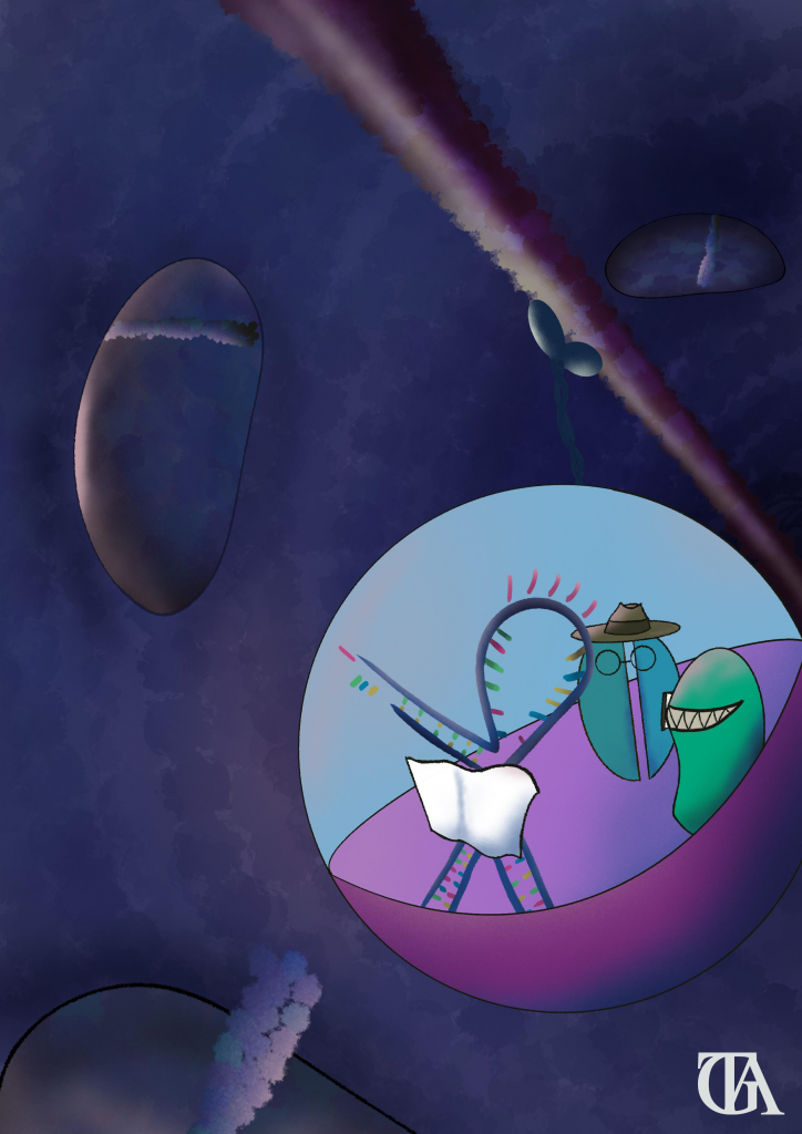
written by Tomás Garnier Artiñano
One might think of cells as unimaginably complex structures made of proteins, sugars, fats, nucleotides, and water (and they are); but they are also much more than that. Around each molecule there is a story; a story of birth, death, adventure, and tragedy. Today, we will follow the story of an adventurous “messenger RNA”; an RNA molecule that acts as the instructions for the production or translation of proteins. This mRNA will venture far from the comfort of its home, the nucleus, and into perilous territories at the fringes of the neurone.
But before we start our journey we must note why our hero is embarking on said adventure. Neurones are cells that communicate information with each other and other cells allowing us to make sense of the world around us. Neurones are highly polarised cells and they require proteins to be at the right place at the right time for connections to form correctly. As an example, memory is a highly complex process where neuronal networks encode for information by changing physical and chemical properties in the neurones, leading to long-term potentiation (LTP) and long term depression (LTD). mRNA localisation is regarded as an important mechanism that fine-tunes protein translation and gene expression. This is very important, as it helps the cell be more energy-efficient and it restricts the effect of a protein to where it is needed, and not where it may cause harm. Overall, mRNA trafficking of neuronal proteins is a complex system that enables neurones to respond accurately to signals from their environment.
As an example, we will follow the journey of the mRNA of the protein CamKII. This is a key protein involved in learning and memory formation. The role of our heroic protein is to induce LTP in the post-synapse. Through in situ hybridisation, the valiant CaMKIIα mRNA has been found localised along the dendrites, the long projections neurones have that receive signals from other neurones, showing that this is one of the brave mRNAs that partakes in the voyage to be translated outside the cell soma. Yet this noble protein has humble beginnings. It is composed of two subunits: CaMKIIα and CaMKIIβ. These two subunits are transcribed by their respective genes in the nucleus of the cell, but while CaMKIIβ gets translated in the soma and then transported to the synapse through conventional protein transport, its sibling, CaMKIIα, has a long and hazardous odyssey ahead.
Just after our mRNA is formed, RNA Binding Proteins (RBP) in the nucleolus bind to the CaMKIIα mRNA molecule. mRNAs contain a molecular zipcode that can target them to different subcellular compartments. It is this zipcode structure that binds to the RBPs to form the ribonuclear particles (mRNPs) that protect the mRNA form the dangerous cytosolic environment and stop the mRNA from producing proteins. Once mRNPs are formed they venture outside the nucleus to continue their journey.
Outside the nucleus, various RNPs clump together or oligomerise and form mRNA transporting granules (TGs). These non-membrane organelles consist of multiple mRNPs bound together into a tight structure. They stop mRNAs from being translated and protect them from cytosolic degradation while delivering its cargo to where it is needed in the cell. Like any respectable party of adventurers, TGs are formed by multiple different mRNAs, not only containing our CaMKIIα, as well as consisting of scaffolding proteins, RNA processing proteins and several parts of the ribosomal translation machinery; all that the CaMKIIα mRNA will need to fulfil its duty once it reaches its destination.
After the granule formation in the neuronal soma, the mRNA team will hitch a ride on the ribo-protein particle. This transporting particle then attaches to the microtubule fibres that run across the neurone and start the passage up the dendrites by using kinesin motors. TGs move up and down the dendrites and can split, combine, and even exchange material with other granules. It is while attached to the cytoskeleton transport machinery that TGs enter the ‘Sushi belt’ mRNA distribution system (and yes, this is the scientific term for it). This model is based on the idea that packaged neuronal mRNAs move bidirectionally along the dendritic cytoskeleton, and are retrieved by nearby synapses when needed.
CaMKIIα mRNA remains on the dendrite transport system until our hero is summoned to action. An action potential stimulus hitting a synapse will act like the bat signal: when this happens, mRNA granules detach from the microtubule filaments and go up the synaptic spines, taking the mRNAs where their presence is required. Synaptic activation loosens the RBP binding partners and allows translation of the delivered mRNA to occur on the synaptic site.
Translation is the last step the CaMKIIα mRNA needs to complete. The TGs are ready for translation as all the necessary machinery can be found inside the mRNA granules. Certain proteins might need special conditions for their correct folding, or to be packaged for secretion. For these situations, fragments of the Endoplasmic Reticulum and the Golgi Apparatus can be found in the dendritic shaft, away from the cell soma. This makes the reaction and adaptation times of neurones many times faster.
As an epilogue to the great quest of CamKIIα, degradation of its mRNA is an inevitable part of the renovation of the mRNA pool. The 3’Cap and PolyA tail are a key factor regulating mRNA stability. Processing bodies (aka, P-bodies) are organelles involved in mRNA degradation and storage. Granules have been observed to exchange material with P-bodies, leading to the cycling of mRNPs. Once inside the P-bodies, enzymes reduce the stability of the mRNA and allow the degradation of the CamKIIα mRNA to happen. And thus the journey of our mRNA comes to an end.
References
Besse, Florence, and Anne Ephrussi. “Translational Control of Localized mRNAs: Restricting Protein Synthesis in Space and Time.” Nature Reviews Molecular Cell Biology, vol. 9, no. 12, 2008, pp. 971–980., doi:10.1038/nrm2548.
Doyle, Michael, and Michael A Kiebler. “Mechanisms of Dendritic MRNA Transport and Its Role in Synaptic Tagging.” The EMBO Journal, vol. 30, no. 17, 2011, pp. 3540–3552., doi:10.1038/emboj. 2011.278.
Glock, Caspar, et al. “mRNA Transport & Local Translation in Neurons.” Current Opinion in Neurobiology, vol. 45, 2017, pp. 169–177., doi:10.1016/j.conb.2017.05.005.
Rangaraju, Vidhya, et al. “Local Translation in Neuronal Compartments: How Local Is Local?” EMBO Reports, vol. 18, no. 5, 2017, pp. 693–711., doi:10.15252/embr.201744045.
